

Breast Cancer is the most common cancer in women worldwide. It occurs when cells located in the breasts divide and grow without control. As it tends to grow slowly, by the time a tumor is large enough to be felt it may have been growing for as long as 10 years. Nowadays there are several procedures to be able to detect breast cancer, nevertheless self-examination should never be forgotten as it is the first line of defense against this illness. A self-exam has to be done once a month and preferably on the same day, just about seven days after your period ends. This helps avoid the hormonal changes that cause texture and consistency changes in the breast and that may cause confusion during the exam.
When doing a self-examination there are some basic steps to follow:
- It should be done in front of a mirror, with your shoulders straight and arms on the hips. You should be searching for any change in size, shape or color. Also that there should be no visible distortion or swelling.
- Then, with arms up, look for the same changes and if there are any signs of fluid coming out of one or both nipples.
- •Next lie down and use your right hand to feel the left breast and your left hand to feel the right breast. Use a circular motion and feel the entire breast side to side, from collarbone to the top of the abdomen and from the arm pit to the cleavage.
There are several procedures to be able to detect breast cancer, nevertheless self-examination should never be forgotten.
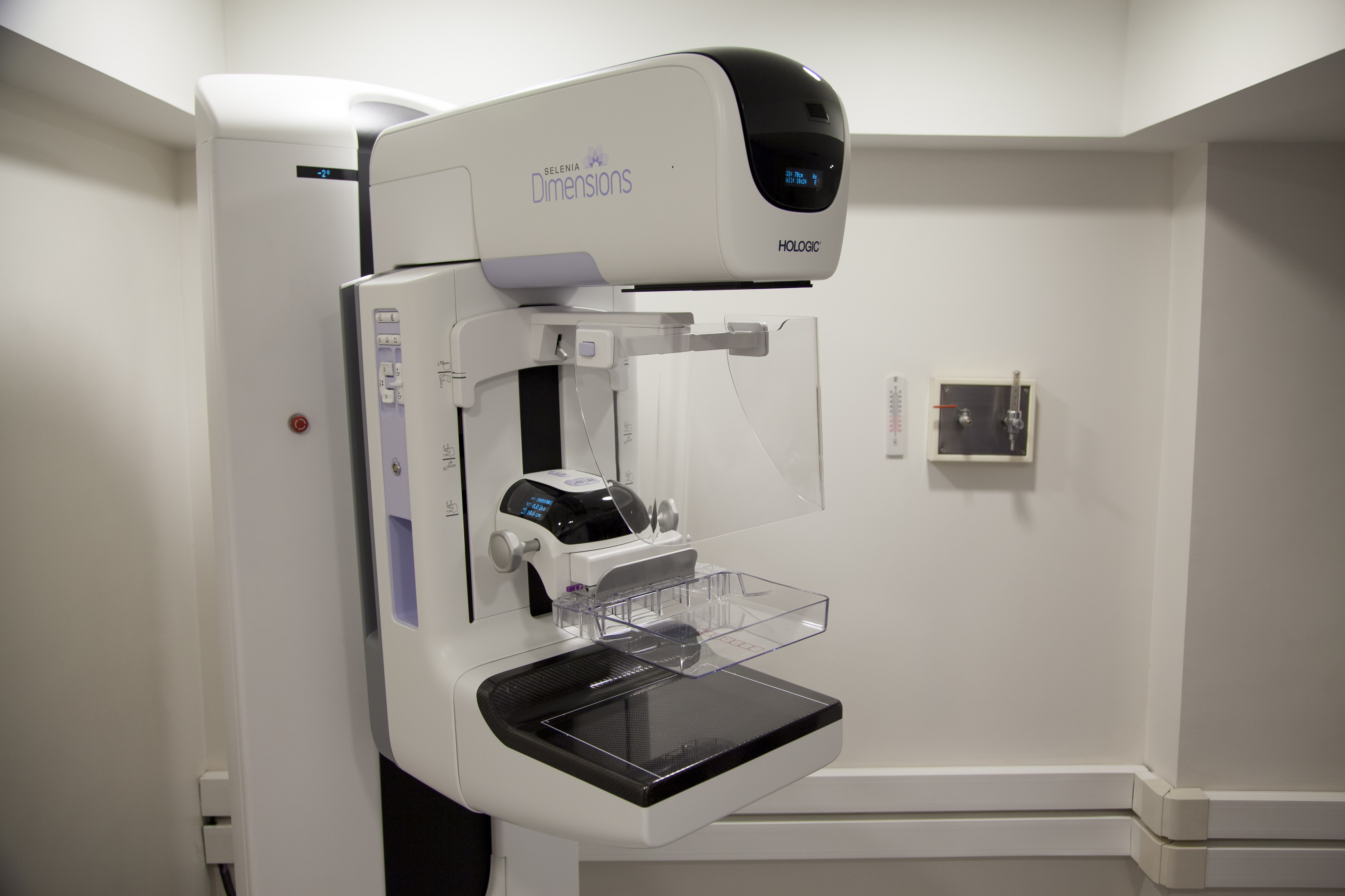
- Then use the same motion to feel your breasts while standing up. If during the self-examination any of the previously noted signs are visible or a lump is felt it should be mentioned to a specialist. Another common procedure is the mammogram which is an X-ray picture of the breast used in women over 40 for early detection. Mammograms can be taken at any hospital or private practice by a specialist. This study should be taken on a yearly basis; the specialist might recommend taking it every six months or every two years, depending on the results of the previous examination. On recent years a new method has become relevant as far as diagnosis and breast cancer stages identification. This is a high definition ultrasound which helps us see and determine each and every one of the lesion characteristics: through a hi-def ultrasound we can diagnose if a tumor is solid or cystic, the tumor’s shape (which is paramount to getting a proper diagnosis), among other factors. This method is generally available, inexpensive and has no health risks. It can be used on women of regardless of age and can be used when a mammogram is not recommended of shows no conclusive results even if discoloration and dense breast tissue appears but a mammogram shows no details of the lesion or tumor. Nowadays technology has turned its way to help medicine, especially with diagnosis of different diseases.
When talking about breast cancer the newest innovation is an ultrasound with elastography, or as some call it “the virtual biopsy”. This helps determine the hardness and elasticity of the lesion and helps physicians decide the most effective course of treatment. Even though this method is not as common as the others, it can be found on some hospitals and specialized private practice. When neither of the above mentioned methods helps in achieving a proper diagnosis, a magnetic resonance imaging (MRI) is used. This method is more expensive than and not as common as the others, but in a particular situation it is extremely helpful.
As with any disease, one of the most important tools we have is information. When having information about a certain condition both of us, patients and doctors, can achieve an early detection which increases the chance of better diagnosis and allows for a better treatment.